Cone Beam – CT (Dental):
Cone-Beam CT images have a higher spatial resolution than conventional CT. Maxilla and mandibula can be scanned in one acquisition (Cylinder: 16 cm. diameter, 13 cm. height), with a substantial lower radiation exposure, and less metal artifacts, compared to classical CT. The images have a 0.2 mm. resolution, in each direction (axial, frontal, sagittal, parasagittal).
Apart from the axial images, 'Dentascan'-type reconstructions can be made if necessary, orthogonal on the dental crest. All images are easily visible through a cloud application, and can be imported in specific dental applications in DICOM format (type Nobelcare, Romexis, ..).
Examples:
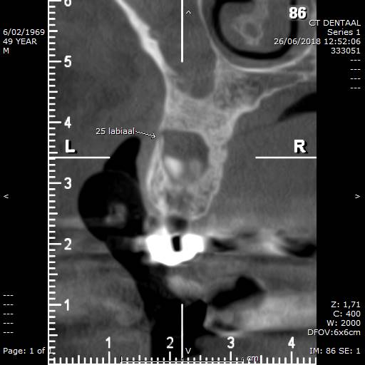
Included and infected molar3, with sinusitis and labial bone perforation.
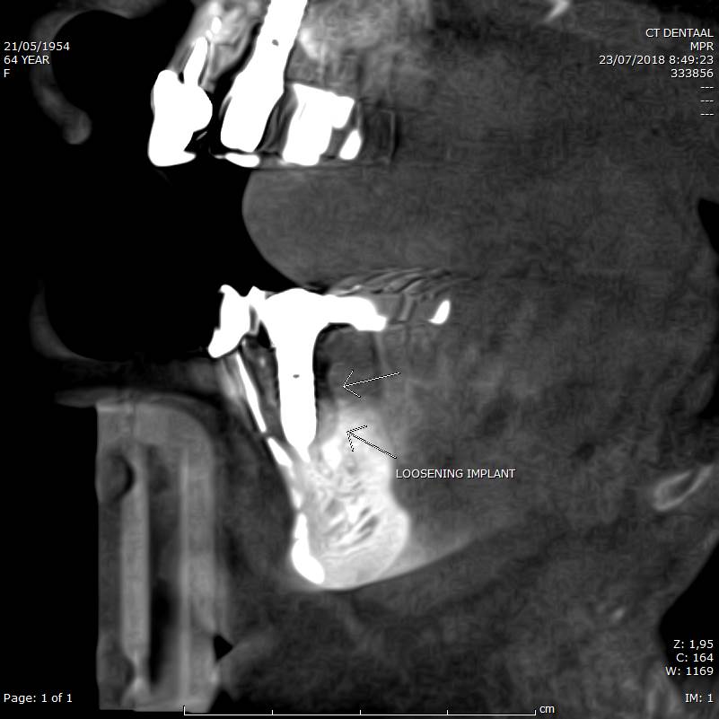
Possible loosening of mandibular implant.
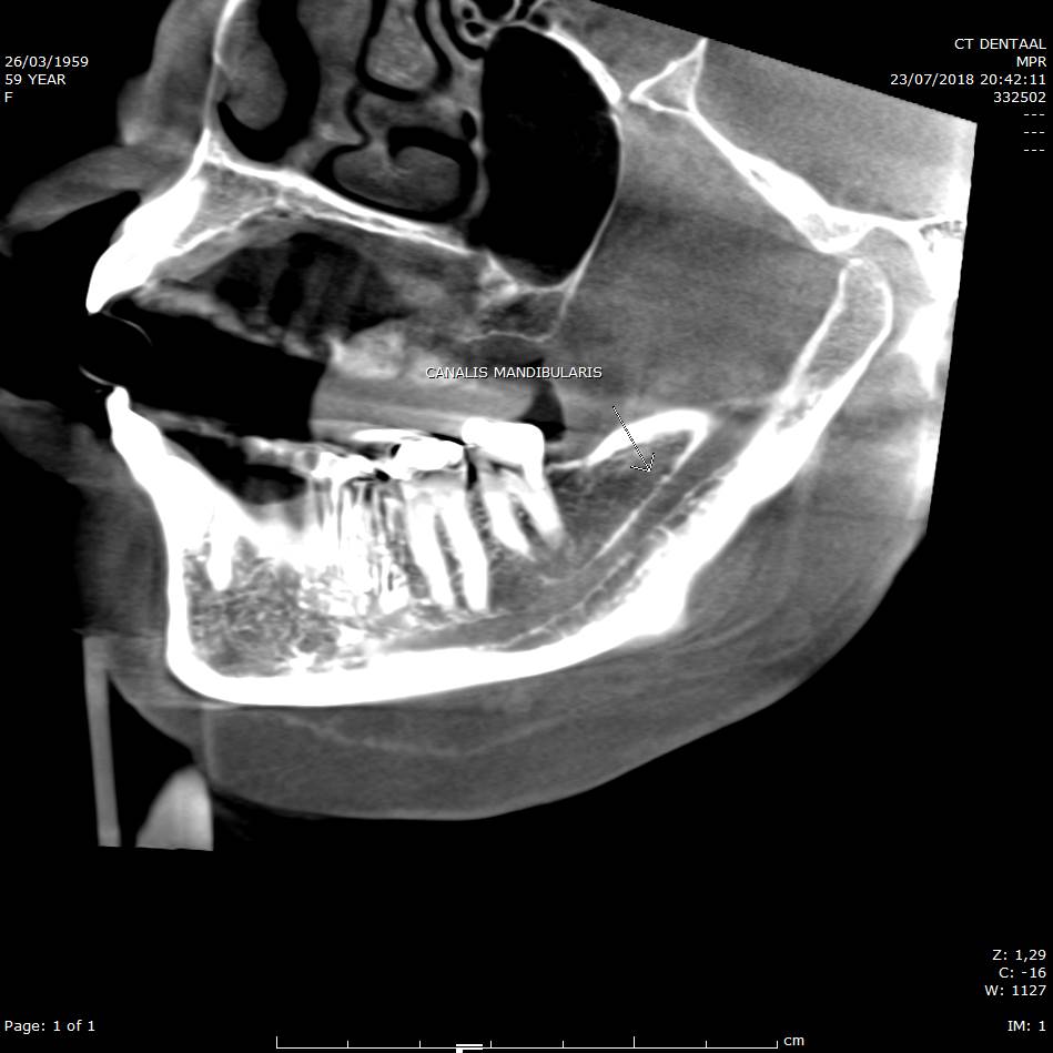
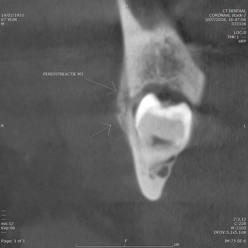
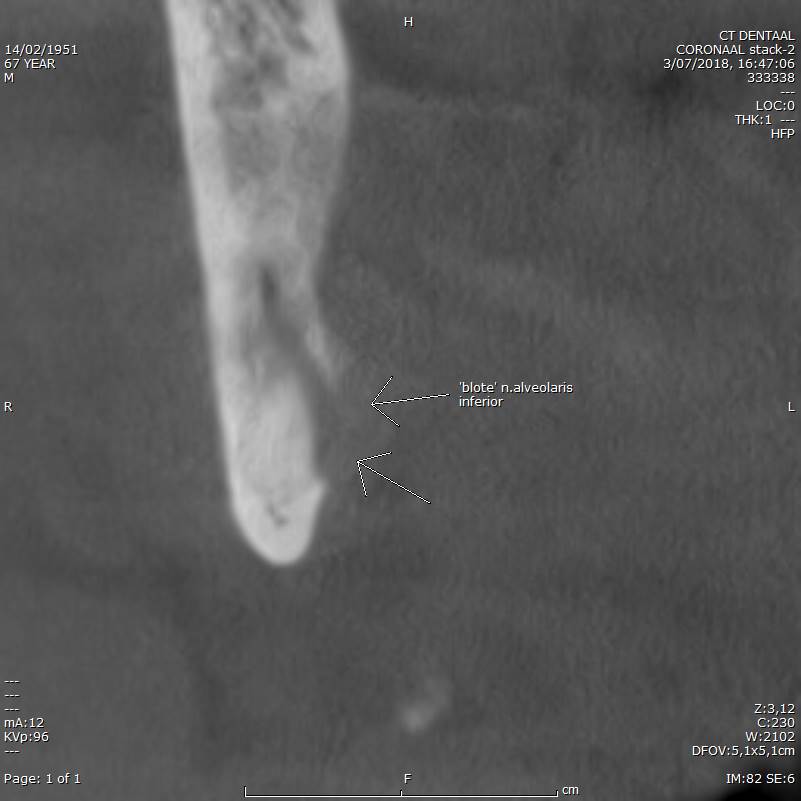
Mandibular M3 infection, with bone perforation, periostal bone apposition, and a bare nervus alveolaris inferior (lingual site).
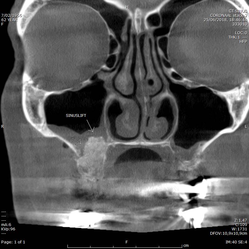
Follow-up after Sinuslift on the right site (even with much metal maxillar and mandibular, still acceptable image quality)

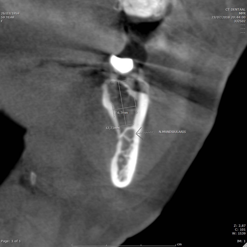
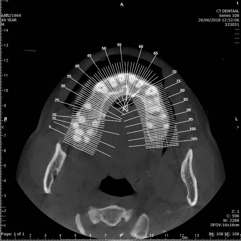
Measurement of bonecrest height and width before implant planning. Location of nervus alveolaris inferior (mandibular nerve).